Diagnostic Procedures
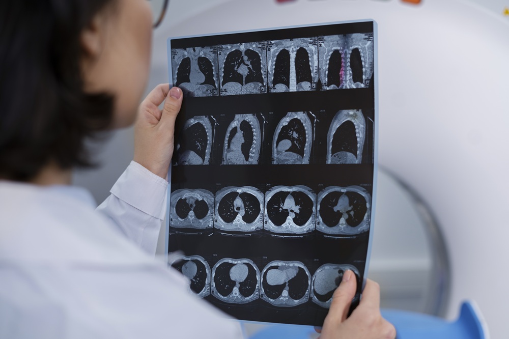
CT Scan
(Computed Tomography)
A rapid imaging technique that uses X-rays and computer processing to create detailed cross-sectional images of the brain, essential for acute conditions.
Duration
5-15 minutes
Contrast Dye
May be used (notify of allergies)
Radiation Dose
Equivalent to 2 yrs background
Description
Computed Tomography (CT) is a rapid imaging technique that uses X-rays and computer processing to create detailed cross-sectional images of the brain. CT is particularly useful for acute conditions requiring rapid diagnosis.
Purpose & Uses
- Acute stroke evaluation (rapid detection of bleeding)
- Traumatic brain injury assessment
- Detection of intracranial hemorrhage
- Evaluation of head trauma
- Detection of brain tumors
- Assessment of sinusitis or skull fractures
- Investigation of sudden severe headache
Advantages
- ✓ Rapid acquisition (minutes)
- ✓ Excellent for detecting bleeding
- ✓ Widely available
- ✓ No contraindications (unlike MRI)
- ✓ Can be performed in emergency settings
Disadvantages
- • Radiation exposure
- • Less soft tissue contrast than MRI
- • Less sensitive for detecting subtle abnormalities
Procedure Steps
CT Scan
- Patient lies on CT table
- Head is positioned in scanner
- Imaging sequences are acquired
- Patient may be asked to hold breath briefly
- Images are reviewed by radiologist
- Patient is moved out of scanner
Results Interpretation
Normal CT
No abnormalities detected.
Abnormal CT
May show bleeding, tumor, fracture, or other abnormalities.
Radiologist provides report with clinical interpretation.
Contrast Dye Information
- • Sometimes used to enhance visualization
- • Injected intravenously
- • May cause warm sensation or metallic taste
- ⚠ Contraindicated in severe kidney disease or iodine allergy
Preparation
- • Remove metal objects
- • Wear comfortable, loose clothing
- • Inform technician of allergies (especially to contrast dye)
- • May require fasting if contrast is used