Carotid Ultrasound
(Carotid Doppler)
A non-invasive imaging test that uses sound waves to visualize the carotid arteries in the neck and assess blood flow.
Duration
15-30 minutes
Discomfort
None; ultrasound is completely painless
Description
Carotid ultrasound is a non-invasive imaging test that uses sound waves to visualize the carotid arteries in the neck and assess blood flow. It helps detect narrowing (stenosis) or plaque buildup that may increase stroke risk.
Purpose & Uses
Procedure Steps
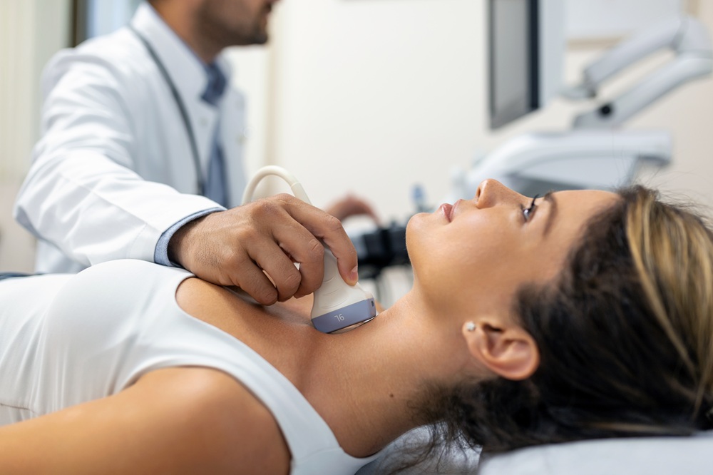
Patient lies on back with head tilted back
Ultrasound gel is applied to neck
Ultrasound probe is moved over carotid arteries
Images and Doppler measurements are obtained
Gel is wiped off
Patient can resume normal activities
Results Interpretation
Normal
No significant stenosis, normal blood flow observed.
Abnormal
Stenosis (mild, moderate, or severe), plaque, or other structural abnormalities detected.
Grading of Stenosis
- Normal: 0% stenosis
- Mild: 1-49% stenosis
- Moderate: 50-69% stenosis
- Severe: 70-99% stenosis
- Occlusion: 100% stenosis
Follow-up Protocols
Repeat ultrasound in 6-12 months
May require intervention (medication or surgery)
May require carotid endarterectomy or stenting
Advantages
- ✓ Non-invasive
- ✓ No radiation
- ✓ No contrast dye needed
- ✓ Rapid and painless
- ✓ Real-time visualization of blood flow
Preparation
- • Wear loose, comfortable clothing
- • No special preparation needed
- • Avoid heavy meals before appointment
Related Conditions
Quick Links
Ready to Schedule Your Test?
Accurate diagnosis is the first step toward effective treatment. Book your Carotid Ultrasound with our specialized neurology team.
Book Carotid Ultrasound