Brain MRI
(Magnetic Resonance Imaging)
A non-invasive imaging technique that uses strong magnetic fields and radio waves to create detailed, high-resolution images of the brain.
Duration
30-60 minutes
Contraindications
Pacemakers, Metal Implants
Safety
Safe, No radiation
Description
Magnetic Resonance Imaging (MRI) is a non-invasive imaging technique that uses strong magnetic fields and radio waves to create detailed, high-resolution images of the brain. MRI provides excellent visualization of brain structures and is particularly useful for detecting soft tissue abnormalities.
Purpose & Uses
- Detection of brain tumors and lesions
- Evaluation of stroke and ischemic changes
- Assessment of multiple sclerosis (MS)
- Investigation of Alzheimer's disease and other dementias
- Evaluation of epilepsy and seizure disorders
- Detection of brain infections (encephalitis, meningitis)
- Assessment of traumatic brain injury
- Evaluation of headaches and migraines
- Detection of vascular abnormalities (aneurysms, AVM)
- Investigation of pituitary disorders
Types of Brain MRI
- Conventional MRI: Standard imaging sequences
- Functional MRI (fMRI): Measures brain activity and blood flow
- Diffusion Weighted (DWI): Detects acute stroke
- Perfusion MRI: Assesses blood flow to brain tissue
- MR Angiography (MRA): Imaging of blood vessels
- MR Spectroscopy: Chemical analysis of brain tissue
Advantages Over CT
- ✓ Superior soft tissue contrast
- ✓ No radiation exposure
- ✓ Better visualization of brain structures
- ✓ Ability to detect subtle abnormalities
- ✓ Multiple imaging sequences available
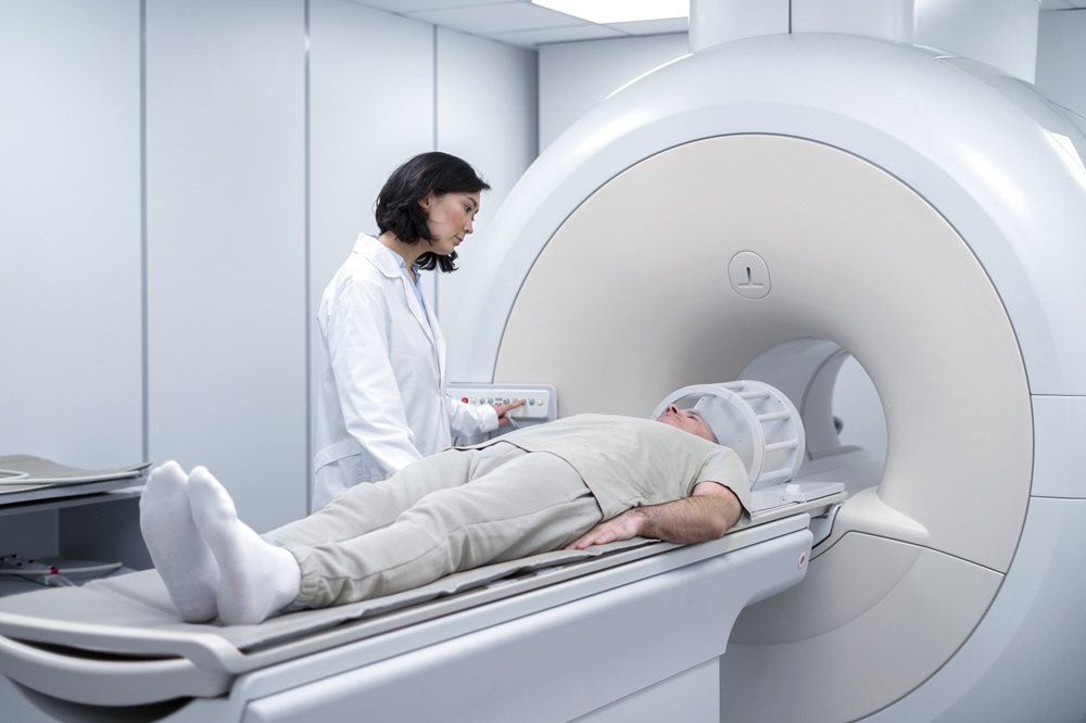
Procedure Steps
- Patient changes into hospital gown (metal-free)
- Patient lies on MRI table
- Safety screening is performed (no metal implants, pacemakers)
- Headcoil is positioned around head
- Table is moved into MRI machine
- Multiple imaging sequences are acquired (20-45 minutes)
- Patient is moved out of machine and images are reviewed
Results Interpretation
Normal MRI
No abnormalities detected in the brain structures or soft tissues.
Abnormal MRI
Findings may include tumors, infarcts, demyelination, infections, or vascular abnormalities.
A Radiologist will provide a detailed report with clinical recommendations.
Contraindications
- • Pacemakers or implantable cardioverter-defibrillators (ICD)
- • Metallic foreign bodies in eyes
- • Ferromagnetic aneurysm clips (older types)
- • Severe claustrophobia (though open MRI may be available)
Preparation
- • Remove all metal objects (jewelry, watches, glasses)
- • Inform technician of any metal implants or medical devices
- • Avoid caffeine before test
- • Wear comfortable, loose clothing
- • Empty bladder before procedure
- • Inform technician if claustrophobic (sedation may be offered)